Saturday, 25 February 2012

PATHOLOGY PRACTICAL # 8 Total Leukocyte Count (TLC) and its interpretation

Total Leukocyte Count (TLC) and its interpretation:

Leukocytes include neutrophils, eosinophils, and basophils, monocytes and lymphocytes.

Granulocytes only include neutrophils, eosinophils, and basophils. While other cells are called agranulocytes such as lymphocytes, monocytes etc.. Neutrophils are the most efficient at phagocytosis and are the principal cells of innate immune defense against bacteria and fungi. 


TLC is recommended to evaluate White cell counting for diagnosing bacterial and viral infections, toxic metabolic processes, diagnosing leukemia stages etc.


                     Normal Adult Total 3500-10,000 cells/cc

CELL TYPE
RANGE (AVG)
Neutrophil
2000-7000 (3700)
Lymphocytes
1500-4000 (2500)
Monocytes
200-1000 (400)
Eosinophils
0-700 (150)
Basophils
0-150 (30)


CLINICAL UTILITY OF CELL COUNTS

CLINICAL CONDITION
DIFFERENTIAL
COUNT FINDING
acute infection
granulocytosis
chronic inflammation
monocytosis
parasitic infection
eosinophilia
viral infection
lymphocytosis
aplastic anemia
neutropenia
acute leukemia
immature cells or
blasts


Marked Leukocytosis can be due to benign or malignant causes:

Peripheral Blood Findings
  (non-malignant)
Chronic Myelogenous Leukemia
WBC > 100000
rare
often
Basophilia
rare
often
Chromosomal abnormalities
never
always
Splenomegaly
rare
often


Eosinophils

Special function of eosinophils is to fight helminths, ticks, and parasites.

Eosinophils can also suppress or enhance hypersensitivity by mobilizing or destroying histamine.

Eosinophils have many red granules.

Eosinophilia is almost always caused by allergic reactions or parasitic infections.  It can cause       tissue destruction.

Basophils

Basophils release histamine and heparin.

They are full of large purple granules.

Basophilia is associated with acute allergic reactions like food allergies.


Monocytes
Monocytes are important for phagocytosis They are also involved in clearing apoptotic cells.
Monocytosis is caused by chronic infection or an inflammatory disease.

Lymphocytes

There are several kinds of lymphocytes (although they all look alike under the microscope), each with different functions to perform . The most common types of lymphocytes are
  • B lymphocytes ("B cells"). These are responsible for making antibodies.
  • T lymphocytes ("T cells"). There are several subsets of these:

Although bone marrow is the ultimate source of lymphocytes, the lymphocytes that will become T cells migrate from the bone marrow to the thymus. where they mature. Both B cells and T cells also take up residence in lymph nodes, the spleen and other tissues where they
  • encounter antigens;
  • continue to divide by mitosis;
  • mature into fully functional cells.


Common Causes of Altered Leukocyte Counts


Decreased
Increased
Neutrophil
Congenital
Hereditary neutropenia

Acquired
Bone marrow disease
Immune reactions
Drugs
Gram-negative septicemia
 
Acquired
Infections
Tissue destruction
Corticosteroids, lithium
Neoplastic growth
Leukemoid reaction, pregnancy,

smoking
Lymphocyte
Congenital
Congenital immunodeficiency disease

Acquired
Severe infection
Drugs (Corticosteroids, alkylating)
GI disease
Immunodeficiency
Acquired
Viral infection (EBV, hepatitis, etc.)
Some fungal, parasitic infections
Rare bacterial infection (Pertussis)
Allergic reactions/drug sensitivities, smoking, CLL
Immunologic disease
Monocyte
Acquired
Hairy cell leukemia
Corticosteroids
Acquired
Mycobacterial infection
Tuberculosis, syphilis
Subacute bacterial endocarditis
Inflammatory responses
Myeloproliferative disorders 
 
Eosinophil
Acquired
Bacterial infection
ACTH administration
Acquired
Parasitic infections, Asthma,
Hypersensitivity reactions
Pulmonary disease
Myeloproliferative diseases
Basophil
Acquired
Corticosteroids
Acquired
Myeloproliferative syndromes
Lymphoproliferative disease
Hypersensitivity reactions
Hodgkin’s disease
Some viral infections
Myxedema


Wednesday, 8 February 2012

Anatomy of the vertebral column and spinal Cord

The spinal cord is the main pathway for information, connecting the brain to the peripheral nervous system.
The human spinal cord is protected by the bony spinal column (vertebral column). The spinal column is made up of bones called vertebrae.

The spinal cord is located in the vertebral foramen and is made up of 31 segments: 8 cervical, 12 thoracic, 5 lumbar, 5 sacral and 1 coccygeal. A pair of spinal nerves exits from each segment of the spinal cord. The spinal cord is about 45 cm long in men and 43 cm long in women.


The Bony Vertebral Column (Spinal Column) 

Ø Supports the head and encloses the spinal cord.
Ø There are 33 bones or vertebrae in the spine:
  § 7 cervical vertebrae in the neck
  § 12 thoracic vertebrae in the upper back corresponding to each
     pair of ribs
  § 5 lumbar vertebrae in the lower back
  § 5 sacral vertebrae are fused together to form 1 bone called
     the sacrum
  § and 4 coccygeal vertebrae that are also fused to form the
     coccyx or tailbone.
Ø We refer to the vertebrae by their name and number so that the
  cervical vertebrae are C1, C2, C3………..C7 where “C” stands for
  “cervical” and the number is the position of the vertebrae counting
  down from the head.
  § Therefore the thoracic vertebrae become T1 –T12
  § The lumbar are L1 – L5
  § And the sacrum and coccyx do not have numbers and each is
     thought of as one bone.

NOTE:

There are 31 pairs of spinal nerves which branch off from the spinal cord. In the cervical region of the spinal cord, the spinal nerves exit above the vertebrae. A change occurs with the C7 vertebra however, where the C8 spinal nerve exits the vertebra below the C7 vertebra. Therefore, there is an 8th cervical spinal nerve even though there is no 8th cervical vertebra. From the 1st thoracic vertebra downwards, all spinal nerves exit below their equivalent numbered vertebrae.


SPINAL NERVES:                                                                   

1.    cervical nerves (nerves in the neck) supply movement and
     feeling to the arms, neck and upper trunk

2.    thoracic nerves (nerves in the upper back) supply the trunk and
     abdomen

3.    lumbar and sacral nerves (from the lower back) supply the legs

How do the vertebrae relate to the spinal cord ?


The level of the spinal cord segments do not relate exactly to the
  level of the vertebral bodies i.e. damage to the bone at a particular
  level e.g. L5 vertebrae does not necessarily mean damage to the
  spinal cord at the same level.

Ø This is because there are:
  § 7 cervical vertebrae but 8 cervical nerve roots leaving the cord
  § the thoracic spinal cord segments are compressed into the level
     between the T1-T10 vertebrae
  § the lumbar and sacral segments are at the level of the T11 and
     T12 vertebrae
  § the spinal cord ends around the level of the disc between T12
     and L1 vertebrae

  § below this level is an extension of the spinal cord called the
     cauda equina (which means “horses tail” in Latin). The cauda
     equina consists of some of the spinal nerves that are continuing
     down before they pass out between the vertebrae.


Saturday, 28 January 2012

Pathology Experiment # 5, 6 and 7

Experiment No. 5
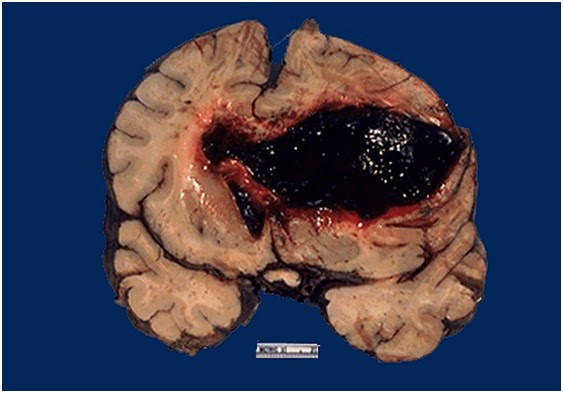
Gross Examination of a hemorrhagic event in the basal ganglia

The large hemorrhage in this adult brain arose in the basal ganglia region of a patient with hypertension. This is one cause for a "stroke".


Experiment # 6
Gross examination of Atrophy and compensatory hypertrophy in kidneys

There is one large-sized kidney with a granular surface and a few scattered, shallow cortical scars. The other kidney shows atrophy because of a narrowed renal artery, most often from atherosclerosis. Such a situation can lead to hypertension.

Experiment # 7
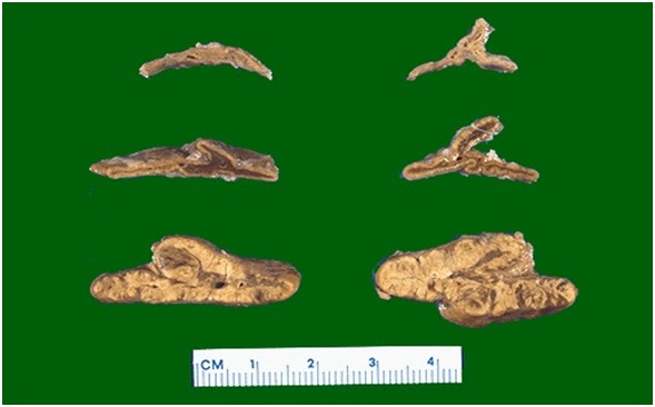
 Gross examination of atrophied and hyperplastic pair of adrenals

The pair of adrenals in the center are normal. Those at the top come from a patient with adrenal atrophy (with either Addison's disease or long-term corticosteroid therapy). The adrenals at the bottom represent bilateral cortical hyperplasia. This could be due to a pituitary adenoma secreting ACTH (Cushing's disease), or Cushing's syndrome from Elevated ACTH production.

Thursday, 5 January 2012

PATHOLOGY Practical No. 2, 3, 4 (Slides + Theory)

Experiment # 2
                       To examine a slide with an arterial atherosclerosis and thrombosis


An atherosclerotic plaque can weaken the wall of an artery, potentially causing a rupture of the vessel; it can cause thrombosis (as it did here) and thereby complete occlusion of the lumen; and it can continue to "grow."

THEORY:
Atherosclerosis often develops at branch points or curving portions along extracranial and intracranial large arteries, locations where blood flow is slowed and more turbulent.  The internal carotid artery is particularly at risk.  Exactly where disease tends to occur, though, and how quickly it progresses apparently reflect the individual's genetic background.  Additional factors that may stimulate plaque growth include hypertension and cigarette smoking.
Atherosclerosis involves focal accumulation of lipid, smooth muscle cells, foamy macrophages, and, eventually, cholesterol crystals under the surface lining (endothelium) of the artery.  With time, such an accumulation can form an elevated plaque that protrudes into the vessel's lumen and significantly reduces blood flow.  Perhaps the analogy of a kitchen drain pipe becoming gradually plugged with cooking grease and sludge will help to visualize what is happening.
If occluding the vascular lumen weren't bad enough, plaques can do a number of additional things that further compromise the brain's circulation.  For instance, a plaque can ulcerate (break open), and the damage to its endothelial lining stimulates the development of a thrombus (blood clot) which even further narrows the vessel.  What's more, pieces of thrombus or fragments of an exposed plaque core can be swept along by the blood flowing through the vessel, becoming emboli.

Atherosclerotic plaques can form in the walls of small arteries as well. However, there are several additional kinds of pathology that particularly affect the walls of small arteries leading to their collapse and blockage of blood flow.
 

Experiment # 3
                  To examine a slide of lung with a pulmonary embolus


THEORY:
Nearly all PEs arise from thrombi in the lower extremity or pelvic veins (deep venous thrombosis [DVT]. Once DVT develops, clots may dislodge and travel through the venous system and right side of the heart to lodge in the pulmonary arteries, where they partially or completely occlude one or more vessels. The consequences depend on the size and number of emboli, the pulmonary reaction, the underlying condition of the lungs, and the ability of the body's intrinsic thrombolytic system to dissolve the clots.

Endogenous lysis reduces most emboli, even those of moderate size, without treatment, and physiologic alterations decrease over hours or days. Some emboli resist lysis and may organize and persist. Occasionally, chronic residual obstruction leads to pulmonary hypertension (chronic thromboembolic pulmonary hypertension) that may develop over years and result in chronic right heart failure. When large emboli occlude major arteries, or when many small emboli occlude > 50% of the distal arterial system, right ventricular pressure increases, causing acute right ventricular failure, failure with shock (massive PE), or sudden death in severe cases. Risk factors for death include age > 70 yr, cancer, and COPD.

Pulmonary infarction occurs in < 10% of patients diagnosed with PE. This low rate has been attributed to the dual blood supply to the lung (ie, bronchial and pulmonary).

PE affects an estimated 117 people per 100,000 person years, resulting in about 350,000 cases yearly, and causes up to 85,000 deaths/yr. PE affects mainly adults.

Symptoms are nonspecific and include dyspnea, chest pain, cough, and, in severe cases, syncope or cardiorespiratory arrest. Signs are also nonspecific and may include tachypnea, tachycardia, hypotension, and a loud pulmonic component of the 2nd heart sound. Diagnosis is based on a CT angiogram, ventilation/perfusion scan, or a pulmonary arteriogram. Treatment is with anticoagulants and, sometimes, clot dissolution with thrombolytics or surgical removal.
                      

Experiment # 4

                     To examine a slide showing Myocardial infarction

Here the infarct shows areas typical coagulative necrosis that typifies anoxic injury. The cells eventually lose their nuclei, lyse and are removed by scavenger cells such as monocytes. All this happened due to an ischemic event in the coronary arteries.
THEORY:
. Myocardial infarction A heart attack or acute myocardial infarction (MI) occurs when one of the arteries that supplies the heart muscle becomes blocked. Blockage may be caused by spasm of the artery or by atherosclerosis with acute clot formation. The blockage results in damaged tissue and a permanent loss of contraction of this portion of the heart muscle.

"Myocardial infarction (MI) is the irreversible necrosis of heart muscle secondary to prolonged ischemia. This usually results from an imbalance of oxygen supply and demand."

Before 6 to 12 hours: No visible lesion is seen. By 18 to 24 hours: Infarct area becomes pale to cyanotic & swollen. In the first week: The infarct area becomes progressively more sharply defined, yellow and softened. By the 7 to 10 days, circumference of the infarct area becomes hyperemic, and progressively expands. By the 6 weeks, fibrous scar is well established.

Electron microscopy shows reversible changes (swelling of mitochondria & endoplasmic reticulum and relaxation of myofibrils).   Histochemically, there is loss of oxidative enzyme & fall of glycogen. In 12 to 72 hours , there is infiltration of neutrophils with progressive coagulative necrosis of myocytes. Dead myocytes become hypereosinophilic with loss of nuclei.